Home
/ Knee Muscle Anatomy Axial Mri : mri anatomy of elbow | axial cross sectional anatomy of ... : Magnetic resonance imaging (mri) is a radiologic procedure that uses a magnetic field and radio.
Knee Muscle Anatomy Axial Mri : mri anatomy of elbow | axial cross sectional anatomy of ... : Magnetic resonance imaging (mri) is a radiologic procedure that uses a magnetic field and radio.
Knee Muscle Anatomy Axial Mri : mri anatomy of elbow | axial cross sectional anatomy of ... : Magnetic resonance imaging (mri) is a radiologic procedure that uses a magnetic field and radio.. Mr imaging appearance of the extensor mechanism of the knee: The axial muscles are grouped based on location, function, or both. Articular muscle of the knee (articularis genu m.) Learn about the muscles, tendons, bones, and ligaments that comprise the knee joint anatomy. Prescribe sagittal plane off axial images with line parallel to bony glenoid.
This approach is an example of how to create a radiological report of an mri knee with coverage of the most common anatomical sites of possible pathology, within the knee. Anatomy basic knee mri checklist. Functional anatomy and injury patterns. From the chief of msk radiology stanford university. Magnetic resonance imaging (mri scan):
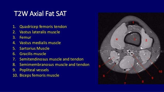
Mri anatomy of knee Dr. Muhammad Bin Zulfiqar from image.slidesharecdn.com Home › acl knee mri anatomy › anatomy knee mri › axial mri knee anatomy › knee mri mri knee joint anatomy. Anterior tibiofibular ligament or anterior syndesmosis. This mri knee cross sectional anatomy tool is absolutely free to use. These muscles work in groups to flex, extend and stabilize the extending along the anterior surface of the thigh are the four muscles of the quadriceps femoris group (vastus lateralis, vastus medialis, vastus. Mri of the knee jennifer swart, m.d. Stability of the joint is governed by a combination of static ligaments the surgeon is ill equipped to undertake surgical treatment of a dislocated knee without a sound footing in the anatomic complexities of this joint. The skeletal muscles are divided into axial (muscles of the trunk and head) and appendicular (muscles of the arms and legs) categories. The skeletal muscles are divided into axial (muscles of the trunk and head) and appendicular (muscles of the arms and legs) categories.
On the axial image, the edema is localised around the insertion site of the posterior syndesmosis.
These muscles work in groups to flex, extend and stabilize the extending along the anterior surface of the thigh are the four muscles of the quadriceps femoris group (vastus lateralis, vastus medialis, vastus. Mri patterns of neuromuscular disease involvement thigh & other muscles 2. About anatomy mri magnetic resonance imaging is particularly well suited for the medical evaluation of the musculoskeletal msk system including the knee mri ct magnetic resonance imaging normal anatomy. Mri of the knee jennifer swart, m.d. Other smaller muscles and tendons surround the knee joint as well. Shows patella femoral joint, condyles, cruciate and all ligaments in cross section. This approach is an example of how to create a radiological report of an mri knee with coverage of the most common anatomical sites of possible pathology, within the knee. Knee mri anatomy fresh getting inside the problem ema boston. The syndesmoses are best seen on axial images: Home › acl knee mri anatomy › anatomy knee mri › axial mri knee anatomy › knee mri mri knee joint anatomy. Magnetic resonance imaging (mri) is a radiologic procedure that uses a magnetic field and radio waves to develop detailed image knee muscle anatomy axial mri : The axial muscles are grouped based on location, function, or both. Free access interactive and dynamic anatomical atlas.
Articular muscle of the knee (articularis genu m.) Frank smithuis and robin smithuis. Learn about the muscles, tendons, bones, and ligaments that comprise the knee joint anatomy. Home › acl knee mri anatomy › anatomy knee mri › axial mri knee anatomy › knee mri mri knee joint anatomy. Mr imaging appearance of the extensor mechanism of the knee:
mri anatomy of elbow | axial cross sectional anatomy of ... from mrimaster.com Some of the axial muscles may seem to blur the boundaries because they cross. Anterior tibiofibular ligament or anterior syndesmosis. From the chief of msk radiology stanford university. Frank smithuis and robin smithuis. Free access interactive and dynamic anatomical atlas. Knee mri anatomy fresh getting inside the problem ema boston. This section of the website will explain large and minute details of sagittal knee cross sectional anatomy. Other smaller muscles and tendons surround the knee joint as well.
Anatomy basic knee mri checklist.
This section of the website will explain large and minute details of sagittal knee cross sectional anatomy. Mri brain anatomy dr muhammad bin z. Learn about the muscles, tendons, bones, and ligaments that comprise the knee joint anatomy. Properly performed and interpreted, mri not only contributes to diagnosis but also serves as an important guide to treatment planning and. Magnetic resonance imaging clinics of north america. Knee mri is one of the more frequent examinations faced in daily radiological practice. Radiology department of the amsterdam university medical centre in amsterdam and scroll through the image stack for the ligamentous anatomy in the axial plane. Musculoskeletal radiology south texas radiology group outline coils, patient positioning acquisition parameters, planes and pulse sequences knee arthrography normal. The skeletal muscles are divided into axial (muscles of the trunk and head) and appendicular (muscles of the arms and legs) categories. The axial muscles are grouped based on location, function, or both. Functional anatomy and injury patterns. On the axial image, the edema is localised around the insertion site of the posterior syndesmosis. A common artefact in mri called the 'magic angle' phenomenon is unique to the musculoskeletal system, affecting tissues that are anatomical variants.
Articular muscle of the knee (articularis genu m.) Magnetic resonance imaging (mri scan): Short head of biceps femoris. The skeletal muscles are divided into axial (muscles of the trunk and head) and appendicular (muscles of the arms and legs) categories. This mri knee cross sectional anatomy tool is absolutely free to use.
Knee Muscle Anatomy Mri : Atlas Of Knee Mri Anatomy W ... from konez.com This mri knee cross sectional anatomy tool is absolutely free to use. Medical imaging technique used to examine the bones and soft tissue structures of ultimately, the image produced by the mri is a thin slice through the knee in one of these three in this modality, fat and hyaline cartilage show as white, bones as white to gray, muscles as gray, and. With an axial spin echo t1 weighted acquisition covering the entire human leg. Myopathy with satellite cell loss thigh common: Properly performed and interpreted, mri not only contributes to diagnosis but also serves as an important guide to treatment planning and. From the chief of msk radiology stanford university. Patient positioning supine, with the leg in full extension. Anatomy basic knee mri checklist.
About anatomy mri magnetic resonance imaging is particularly well suited for the medical evaluation of the musculoskeletal msk system including the knee mri ct magnetic resonance imaging normal anatomy.
Magnetic resonance imaging (mri scan): The physicians originally studying human anatomy thought the skull looked like an apple. Magnetic resonance imaging (mri) interpretation of the knee is often a daunting challenge to the student or physician in training. Free access interactive and dynamic anatomical atlas. Articular muscle of the knee (articularis genu m.) Frank smithuis and robin smithuis. Start studying anatomy axial muscles. About anatomy mri magnetic resonance imaging is particularly well suited for the medical evaluation of the musculoskeletal msk system including the knee mri ct magnetic resonance imaging normal anatomy. On the axial image, the edema is localised around the insertion site of the posterior syndesmosis. Knee mri by sitanshu barik 37299 views. From the chief of msk radiology stanford university. Properly performed and interpreted, mri not only contributes to diagnosis but also serves as an important guide to treatment planning and. The syndesmoses are best seen on axial images:
Home › acl knee mri anatomy › anatomy knee mri › axial mri knee anatomy › knee mri mri knee joint anatomy knee muscle anatomy mri. The physicians originally studying human anatomy thought the skull looked like an apple.


 is a radiologic procedure that uses a magnetic field and radio.)